
Doppler Test in Pokhara — Colour Doppler Ultrasound at KDRC
A Doppler test in Pokhara at KDRC is a colour Doppler ultrasound scan that measures the speed and direction of blood flow in arteries and veins. It detects clots, narrowed or blocked vessels, varicose veins and reduced blood supply to organs or the baby, with no radiation, no needles and usually no fasting.
Also called: colour doppler, doppler test, duplex ultrasound, color doppler, doppler scan, venous doppler
 Doppler UltrasoundKDRC · Pokhara
- Duration
- Most Doppler studies take about 15 to 45 minutes,
- Reported by
- Consultant radiologists
- Location
- Amarsingh Chowk, Pokhara-10
- Book / price
- 061553538 · 9856077433
- Doppler ultrasound measures blood flow, which a standard ultrasound image alone cannot show.
- It is the usual first test for leg clots (DVT), varicose veins, leg artery disease and neck artery narrowing.
- It uses no radiation and no contrast, so it is safe in pregnancy when indicated, in children and in kidney disease.
- Most studies need no fasting; abdominal and kidney Doppler may need it, and staff confirm when you book.
- KDRC Doppler studies are reported by consultant radiologists, with CT angiography and MRI on site for next steps.
On this page (14 sections)
- What is a Doppler test?
- How does Doppler ultrasound work?
- Why is a Doppler test done?
- Venous Doppler in Pokhara: blood clots and varicose veins
- Carotid Doppler: checking the neck arteries
- Arterial Doppler for leg pain, diabetes and poor circulation
- Obstetric Doppler during pregnancy
- How to prepare for a Doppler test
- What happens during a Doppler scan at KDRC?
- Is a Doppler test safe and painful?
- Doppler results and reports
- How much does a Doppler test cost in Pokhara?
- Why choose KDRC for a Doppler test in Pokhara?
- Getting to KDRC and booking your Doppler test
What is a Doppler test?
A Doppler test is an ultrasound scan that measures the speed and direction of blood flowing through your arteries and veins. A Doppler test in Pokhara at KDRC checks blood flow without needles or radiation. A normal ultrasound shows the shape of a vessel. Doppler adds the movement of the blood inside it. Together they show whether a vein holds a clot, whether an artery is narrowed, and whether blood reaches an organ as it should.
Patients and doctors use many names for the same test: Doppler ultrasound, colour Doppler, duplex scan or vascular ultrasound. In Pokhara people often simply ask for a "Doppler test" or a "colour Doppler". Because Doppler is a special mode of ultrasound, some patients also call it a "video X-ray" (भिडियो एक्स-रे), but no X-ray is used at any point. If you are looking for a Doppler test in Pokhara, this page explains what it is, why your doctor asked for it, how to prepare, and what happens at Kaligandaki Diagnostic and Research Center (KDRC) on Shantipath, Amarsingh Chowk.
How does Doppler ultrasound work?
Doppler ultrasound works by sending sound waves into the body and measuring the small change in pitch when they bounce back from moving red blood cells. This change is called the Doppler effect. It is the same effect that makes a passing motorbike sound higher as it approaches and lower as it moves away. Blood moving towards the probe returns a slightly higher frequency. Blood moving away returns a lower one. The machine turns these changes into colours, graphs and sound.
During the scan you will often hear a rhythmic "whooshing" noise. That is the Doppler signal of your own blood flow. Arteries usually give a pulsing sound with each heartbeat. Veins usually give a softer, steadier sound that changes with breathing.
Several Doppler modes are combined in one examination, and each answers a slightly different question:
| Doppler mode | What it shows | Typical use |
|---|---|---|
| Colour Doppler | Flow painted in colour over the grey-scale image; colour indicates direction relative to the probe | Quickly finding narrowed segments, clots, reflux and abnormal vessels |
| Spectral (pulsed-wave) Doppler | A graph of blood speed over time at one chosen point | Measuring how tight a narrowing is; judging the pattern of flow |
| Power Doppler | Very sensitive display of the presence of flow, not its direction | Detecting slow or low-volume flow, for example in small organs |
| Duplex scan | Grey-scale image and Doppler flow shown together | Most venous and arterial studies of the legs, arms and neck |
Red and blue on the screen do not mean "artery" and "vein". They only show whether blood is moving towards or away from the probe. The radiologist interprets the colours together with the waveforms and the grey-scale picture.
Why is a Doppler test done?
A Doppler test is done to find problems with blood flow, such as clots, narrowed or blocked vessels, leaking vein valves and reduced blood supply to an organ. RadiologyInfo lists its main uses as locating blockages and plaque, detecting deep vein clots, finding aneurysms and varicose veins, checking grafts and bypasses, and showing reduced or increased blood flow to organs. The study your doctor requests depends on your symptoms. The table below matches common Doppler studies to the questions they answer.
| Doppler study | Vessels examined | Common reasons for referral |
|---|---|---|
| Venous Doppler (lower or upper limb) | Deep and superficial veins of the leg or arm | One-sided swelling or pain, suspected deep vein thrombosis (DVT), varicose veins, leg ulcers |
| Arterial Doppler (lower limb) | Arteries from the groin to the foot | Leg pain on walking, cold feet, non-healing wounds, diabetes, smoking-related artery disease |
| Carotid Doppler | Carotid and vertebral arteries in the neck | Stroke or mini-stroke (TIA) assessment, a neck bruit, high stroke risk |
| Abdominal and renal Doppler | Aorta, kidney arteries, liver and portal veins | Aneurysm, high blood pressure work-up, liver disease, kidney assessment |
| Obstetric Doppler | Umbilical cord and, when needed, fetal or placental vessels | Checking blood flow to the baby when the doctor has a clinical concern |
| Scrotal or pelvic Doppler | Blood supply to the testes or ovaries | Sudden pain, swelling, suspected reduced or absent blood flow |
Some of these reasons can be emergencies. Please read the warning below before booking a routine appointment.
Go to a hospital emergency department immediately if leg swelling or pain comes with chest pain or sudden breathlessness, which can signal a clot in the lungs. Also seek emergency care for sudden face drooping, arm weakness or slurred speech, a cold, pale or numb leg, or sudden severe testicular pain. Call an ambulance, such as the 24/7 KDRC ambulance; do not drive yourself.
Venous Doppler in Pokhara: blood clots and varicose veins
A venous Doppler checks the veins for clots and for valves that let blood flow backwards. It is the most common Doppler study requested in everyday practice.
Deep vein thrombosis (DVT)
Deep vein thrombosis is a blood clot in a deep vein, usually in the calf or thigh. The NHS describes typical symptoms as throbbing pain and swelling in one leg, warm or discoloured skin, and swollen veins. NHS guidance says that when a doctor suspects DVT, the patient should have an ultrasound scan within 24 hours to check whether blood is flowing normally through the vein. Risk rises after surgery, during a hospital stay, with bed rest of 3 days or more, on long journeys of over 4 hours, during pregnancy and in the 6 weeks after giving birth. A venous Doppler looks along the deep veins, presses gently on them with the probe and checks the flow. A healthy vein squeezes flat; a vein containing a clot does not.
Varicose veins and venous reflux
Varicose veins are swollen, twisted veins, usually in the legs. The NHS notes that a specialist usually confirms them with a duplex ultrasound, which checks blood flow and helps plan treatment. During a venous reflux study you may be asked to stand or sit with your legs down, take a deep breath and strain, or have your calf squeezed. These manoeuvres show whether the vein valves close properly. The resulting map tells the surgeon which veins are leaking before any procedure.
Carotid Doppler: checking the neck arteries
A carotid Doppler examines the two large arteries in the neck that carry blood to the brain, looking for narrowing (stenosis) that can raise the risk of stroke. RadiologyInfo lists its uses as screening for narrowing or blockage, assessing patients with high blood pressure, diabetes, high cholesterol, a neck bruit or a family history of stroke or heart disease, and checking an artery after surgery or stenting. You lie on your back with your head turned slightly away from the side being scanned. An open-necked shirt or kurta makes the scan easier. The radiologist measures the speed of blood through any narrowing, because faster flow suggests a tighter stenosis. Carotid Doppler does not replace emergency stroke care: anyone with sudden face drooping, arm weakness or slurred speech needs a hospital emergency department straight away.
Arterial Doppler for leg pain, diabetes and poor circulation
An arterial Doppler of the legs looks for narrowed or blocked arteries that reduce blood supply to the muscles and skin. The commonest symptom of peripheral arterial disease is leg pain when walking that eases after a few minutes of rest, known as intermittent claudication. The NHS names smoking as the most significant risk factor, followed by diabetes, high blood pressure, high cholesterol and older age. Many people have no symptoms at all.
The study often begins at the groin and follows the arteries down to the ankle. The radiologist notes where flow changes and how severe any narrowing appears. For patients with diabetes, Doppler findings help doctors decide how to look after foot wounds. If the doctor needs a full road map of the arteries before surgery or angioplasty, Doppler is often followed by CT angiography at KDRC, which shows all the vessels at once using contrast.
Obstetric Doppler during pregnancy
Obstetric Doppler measures blood flow in the umbilical cord and, when needed, in the baby or placenta, and it is requested when a doctor wants extra information about the baby's well-being. RadiologyInfo notes that Doppler may be part of a pregnancy ultrasound and that ultrasound has been used in pregnancy for nearly four decades with no evidence of harm to the mother, embryo or fetus. It should still be done only when medically indicated.
Doctors usually request obstetric Doppler when they are concerned about a baby's growth, the amount of fluid around the baby, or the mother's health. It is a focused add-on rather than a routine screening scan. The detailed structural check of the baby is a separate examination: see our anomaly scan in Pokhara page for that, and the HD ultrasound page for dating, growth and 3D/4D scans. If your doctor requests a growth scan with Doppler, our team can usually perform both in the same visit on the same machine.
How to prepare for a Doppler test
Most Doppler tests need no special preparation beyond comfortable clothing, but a few abdominal studies need fasting. RadiologyInfo advises leaving jewellery at home and wearing loose clothing, and says your doctor will tell you if you should avoid eating or drinking. Our booking staff confirm your exact instructions when you call. The table gives the usual preparation by study type.
| Study | Food and drink | What to wear and bring |
|---|---|---|
| Leg or arm venous Doppler | Eat and drink normally | Loose trousers or a skirt; shorts can help for leg scans |
| Leg arterial Doppler | Eat and drink normally; avoid smoking just before | Loose clothing; a list of diabetes and heart medicines |
| Carotid Doppler | Eat and drink normally | Open-necked top; remove necklaces |
| Abdominal or renal Doppler | Fasting is often requested; staff will confirm the hours | Two-piece outfit; previous abdominal reports |
| Obstetric Doppler | Eat and drink normally unless told otherwise | Two-piece outfit; antenatal card and earlier pregnancy scans |
- Doctor's request form naming the vessels to be scanned
- Citizenship card or other ID
- List of current medicines, especially blood thinners and diabetes medicines
- Previous Doppler, CT angiography or surgery reports
- Loose clothing or a two-piece outfit; leave jewellery at home
- Avoid smoking just before an arterial or carotid study
- Ask staff whether fasting is needed for an abdominal or renal Doppler
What happens during a Doppler scan at KDRC?
A Doppler scan at KDRC is a painless, hands-on ultrasound examination that usually takes between a quarter of an hour and three quarters of an hour. The NHS gives 10 to 45 minutes as the typical range for an ultrasound scan, and a full study of both legs or of arteries and veins together can take longer. The steps below describe a typical visit from registration to report.
- 1Register at receptionShow your request form and ID at the Shantipath reception. Staff confirm the study and any preparation.
- 2Change and positionYou uncover the limb, neck or abdomen to be scanned and lie on the couch. For some venous studies you sit or stand.
- 3Gel and probeWarm gel is applied and the probe is moved along the vessels. Colour and spectral Doppler measure the flow.
- 4ManoeuvresYou may be asked to breathe deeply, strain, or have your calf squeezed so that valve function can be tested.
- 5Finish and clean upThe gel is wiped off. You can eat, drive and return to work immediately.
- 6ReportThe consultant radiologist reviews the images and issues the report. Staff tell you when to collect it.
You can talk during most of the scan. The sonographer or radiologist may ask you to hold your breath, turn slightly, or keep very still for a few seconds while a measurement is recorded. Firm probe pressure over a tender, swollen calf can be uncomfortable for a moment; tell the examiner and they will adjust.
Is a Doppler test safe and painful?
A Doppler test is safe and is not painful, because it uses sound waves rather than radiation and involves no injections. RadiologyInfo states that vascular ultrasound is non-invasive, uses no ionising radiation and has no known harmful effects when used properly. Mild pressure is the only common discomfort. It is safe in pregnancy when medically indicated, safe for children, and safe for people with kidney disease, because no contrast dye is needed.
Doppler does have limits. Deep vessels in larger patients, very small vessels and arteries heavily coated with calcium can be hard to see, and image quality depends on the skill of the examiner. When Doppler cannot answer the question, doctors may move on to CT angiography or MRI. The comparison below shows how Doppler sits next to CT angiography.
| Feature | Doppler ultrasound | CT angiography |
|---|---|---|
| Radiation | None | Yes, X-ray based |
| Contrast injection | Not needed | Iodine contrast through a vein |
| Pregnancy | Safe when medically indicated | Usually avoided unless essential |
| Shows real-time flow | Yes, with direction and speed | Shows vessel anatomy at one moment |
| Deep or calcified vessels | Can be hard to see | Shows them well |
| Best first test for DVT and varicose veins | Yes | Rarely needed |
Doppler results and reports
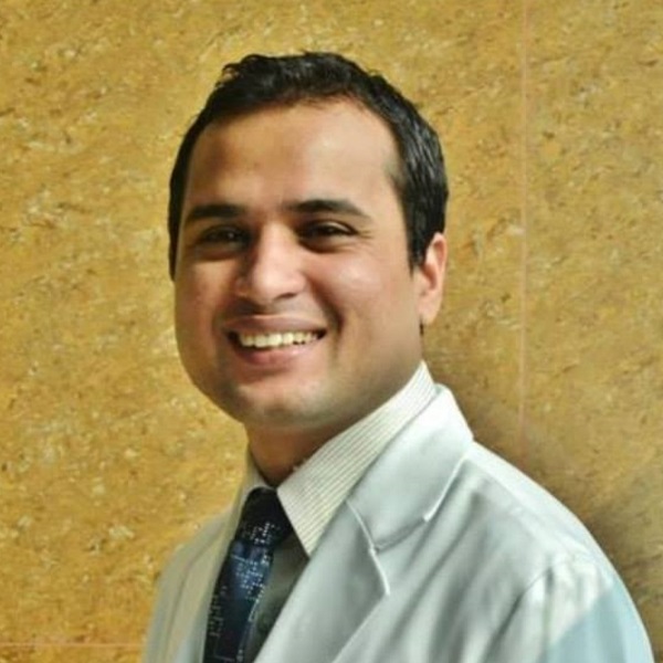
Your Doppler report describes each vessel examined, the direction and speed of flow, and any clot, narrowing or reflux the radiologist found. At KDRC, Doppler studies are reported by our consultant radiologists, Dr. Bishworaj Bastakoti and Dr. Nawaraj Paudel. Both have been registered radiologists with the Nepal Medical Council since 2016 and are members of the Nepal Radiologist Society. Dr. Bastakoti trained at the National Kidney and Transplant Institute in Manila and is a Diplomate of the Philippine College of Radiology. Dr. Paudel trained at East Avenue Medical Centre in Manila and is a former assistant professor of radiology at Universal College of Medical Sciences.
Report timing depends on the study and how complex the findings are. Our team will tell you when to collect your report. If a finding needs urgent attention, such as a fresh deep vein clot, the radiologist will advise you to see your doctor without delay. Please bring earlier Doppler reports, because comparing old and new measurements shows whether a narrowing or reflux is stable or getting worse. Referring hospitals that need remote reporting can also use our teleradiology service.
How much does a Doppler test cost in Pokhara?
The cost of a Doppler test in Pokhara depends mainly on which vessels are examined and how many, so KDRC quotes each study individually. A single-leg venous scan is a smaller study than both legs, and both are smaller than a combined arterial and venous examination. A carotid Doppler, a renal Doppler and an obstetric Doppler are each priced separately. If Doppler is added to another ultrasound in the same visit, staff can explain how that affects the total. For the current Doppler price, call 061-553538 or 9856077433 (also on Viber) and tell us which study your doctor has written on the request form.
Why choose KDRC for a Doppler test in Pokhara?
KDRC combines high-definition colour Doppler with consultant radiologist reporting and a full imaging department under one roof, so a Doppler result can lead straight to the next test without travelling across the city.
- HD ultrasound with colour Doppler: the same high-definition system used for our 3D, 4D and volume scans.
- Consultant radiologists: two NMC-registered radiologists who have worked at KDRC since the centre began.
- Next steps on site: CT angiography on a 192-slice Siemens CT, open-type MRI and ultrasound-guided FNAC when a Doppler finding needs further work-up.
- Help with transport: a 24/7 ambulance service for patients who cannot travel safely.
- Any day of the week: ultrasound-based services, including anomaly scans, are available on any day of the week; call to confirm a time.
- Same platform; structure of organs
- Next step for a full vessel map
- Ultrasound for patients who cannot travel
- Structural pregnancy scan; Doppler can be added
- Further imaging when ultrasound is limited
- Transport for urgent cases
Getting to KDRC and booking your Doppler test
KDRC is on Shantipath at Amarsingh Chowk, Pokhara-10, Kaski. Most local taxi drivers know Amarsingh Chowk, and the centre's signage is visible from the road. Bring your doctor's request form, your citizenship card or another ID, a list of your medicines and any earlier vascular reports. If you are coming from outside Pokhara, for example from Syangja, Tanahun, Parbat or Baglung, call first so that the study can be confirmed before you travel.

To book, call 061-553538 or 9856077433, email kaligandaki.dc@gmail.com, or use the appointment form. You can browse all KDRC services if your doctor has requested more than one test. Ask whether a referral is needed for your particular study; staff will guide you.
How to prepare
- Bring your doctor's request form, ID, medicine list and earlier vascular reports.
- Wear loose clothing or a two-piece outfit; leave jewellery at home.
- Eat and drink normally for limb, neck and pregnancy Doppler studies.
- Ask whether fasting is needed for abdominal or kidney Doppler.
- Avoid smoking just before an arterial or carotid study.
- Keep taking your usual medicines unless your doctor says otherwise.
How long it takes
Most Doppler studies take about 15 to 45 minutes, and combined studies of both legs or of arteries and veins can take longer.
Price in Pokhara
Doppler cost depends on which vessels are examined and how many, so each study is quoted individually. Call 061-553538 or 9856077433 for the current price.
What to expect at KDRC
When you arrive at our Shantipath centre, show your doctor's request form and ID at reception. Staff confirm which vessels are being studied and check any preparation, such as fasting for an abdominal Doppler. In the scanning room you uncover the area to be examined and lie on the couch; for some vein studies you sit or stand so the veins fill. Warm gel goes on the skin and the probe is moved slowly along the vessels while colour and spectral Doppler record the flow. You will hear a whooshing sound, which is your own blood moving. You may be asked to breathe deeply, strain, or have your calf gently squeezed. Most studies take between about fifteen and forty-five minutes, longer if both legs or both arteries and veins are examined. Afterwards the gel is wiped away and you can eat, drive and go back to work straight away. A consultant radiologist reviews the images and writes the report, and our team tells you when to collect it for your doctor.
Frequently asked questions
Where can I get a Doppler test in Pokhara?
You can get a Doppler test at Kaligandaki Diagnostic and Research Center (KDRC), Shantipath, Amarsingh Chowk, Pokhara-10. We perform venous, arterial, carotid, abdominal and obstetric Doppler studies on a high-definition ultrasound system with colour Doppler, reported by consultant radiologists. Call 061-553538 or 9856077433 to book a time.
What is the difference between ultrasound and Doppler?
A standard ultrasound shows the shape and structure of organs and vessels. Doppler is an extra mode of the same machine that measures the speed and direction of blood flow. Many scans combine both, which is called a duplex scan. Your doctor's request form will say whether Doppler is needed.
What is colour Doppler?
Colour Doppler is a Doppler mode that paints blood flow in colour on top of the grey-scale ultrasound image. Red and blue show whether blood moves towards or away from the probe, not whether a vessel is an artery or vein. It helps the radiologist quickly find clots, narrowing and leaking valves.
How much does a Doppler test cost in Pokhara?
The cost depends on which vessels are scanned and how many, for example one leg, both legs, arteries and veins together, the neck arteries or an obstetric study. KDRC quotes each study individually and prices change from time to time. Call 061-553538 or 9856077433 with your request form details for the current price.
How long does a Doppler scan take?
Most Doppler scans take roughly 15 to 45 minutes. The NHS gives 10 to 45 minutes as the usual range for ultrasound scans, and a study of both legs, or of arteries and veins together, can take longer. Allow extra time for registration and for waiting on busy days.
Do I need to fast before a Doppler test?
Usually not. Leg, arm, neck and pregnancy Doppler studies need no fasting, so you can eat and drink normally. Abdominal and kidney Doppler studies often need a period of fasting so that bowel gas does not block the view. Our staff confirm the exact instructions when you book.
Is a Doppler test painful?
No. A Doppler test uses a smooth probe and gel on the skin, with no needles or injections. You may feel firm pressure, especially over a swollen or tender leg, and a few manoeuvres such as straining or a calf squeeze can be briefly uncomfortable. Tell the examiner and they will adjust.
Is Doppler safe during pregnancy?
Yes, when it is medically indicated. Doppler uses sound waves, not radiation, and RadiologyInfo notes that ultrasound has been used in pregnancy for nearly four decades with no evidence of harm. Doctors request obstetric Doppler when they need information about blood flow to the baby, not as a routine extra.
Can a Doppler test detect a blood clot in the leg?
Yes. A venous Doppler is the standard first test for a suspected deep vein thrombosis. The examiner presses gently on the veins and checks the flow; a vein containing a clot does not squeeze flat. If you also have chest pain or breathlessness, go to an emergency department immediately instead of booking a routine scan.
Can children have a Doppler test?
Yes. Doppler has no radiation and no injection, so it is suitable for children when a doctor requests it, for example for sudden testicular pain or kidney problems. A parent can stay in the room. Bringing a favourite toy or phone video helps a young child lie still for the measurements.
Do I need a doctor's referral for a Doppler test?
A doctor's request is strongly recommended, because it tells the radiologist which vessels to examine and what question to answer. Doppler covers many different studies, and the right one depends on your symptoms. Call our staff if you are unsure; they will guide you on what your request form should say.
Can I drive or go to work after a Doppler scan?
Yes. A Doppler scan needs no sedation, injection or recovery time, so you can drive, eat and return to work straight afterwards. The gel wipes off easily. The only exception is if the radiologist finds something that needs urgent care, in which case they will advise you to see your doctor promptly.
Who reports Doppler scans at KDRC?
Doppler studies at KDRC are reported by consultant radiologists Dr. Bishworaj Bastakoti and Dr. Nawaraj Paudel. Both have been NMC-registered radiologists since 2016, are members of the Nepal Radiologist Society, and have worked at KDRC since the centre began. Their reports go to your referring doctor with you.
When will I get my Doppler report?
Timing depends on the type of study and how complex the findings are. Our team will tell you when to collect your report at the time of your scan. Urgent findings, such as a new deep vein clot, are flagged so that you can see your doctor without delay. Bring earlier reports for comparison.
Is Doppler better than CT angiography?
Neither is better for every case. Doppler is radiation-free, needs no contrast and shows real-time flow, so it is the usual first test for clots, varicose veins and neck arteries. CT angiography shows deep and calcified vessels better and maps a whole region at once, so doctors use it when surgery is planned.
Sources
- radiologyinfo.org/en/info/vascularus
- radiologyinfo.org/en/info/us-carotid
- radiologyinfo.org/en/info/obstetricus
- nhs.uk/conditions/deep-vein-thrombosis-dvt/
- nhs.uk/conditions/varicose-veins/
- nhs.uk/conditions/peripheral-arterial-disease-pad/
- nhs.uk/tests-and-treatments/ultrasound-scan/
This page is general health information reviewed by our radiologists. It is not a diagnosis — always follow your own doctor's advice. In an emergency, call our 24-hour ambulance or go to the nearest emergency department.
Shantipath, Amarsingh Chowk, Pokhara-10, Kaski · 061553538 / 9856077433
Related services in Pokhara
Open Type MRI
An MRI in Pokhara at KDRC is a scan that uses a strong magnet and radio waves, not X-rays, to picture the brain, spine, …
High Definition Ultrasound
Ultrasound (USG, sonography) in Pokhara at KDRC is a painless, radiation-free scan that uses high-frequency sound waves …
CT Angiography
CT angiography in Pokhara at KDRC is a contrast-enhanced CT scan that maps the arteries and veins of the heart, brain, n…
Mobile Ultrasound
Mobile ultrasound service in Pokhara at KDRC brings a portable ultrasound machine and trained team to the bedside of pat…